14.11: Phosphates, Arsenates, and Vanadates
- Page ID
- 18684
\( \newcommand{\vecs}[1]{\overset { \scriptstyle \rightharpoonup} {\mathbf{#1}} } \)
\( \newcommand{\vecd}[1]{\overset{-\!-\!\rightharpoonup}{\vphantom{a}\smash {#1}}} \)
\( \newcommand{\dsum}{\displaystyle\sum\limits} \)
\( \newcommand{\dint}{\displaystyle\int\limits} \)
\( \newcommand{\dlim}{\displaystyle\lim\limits} \)
\( \newcommand{\id}{\mathrm{id}}\) \( \newcommand{\Span}{\mathrm{span}}\)
( \newcommand{\kernel}{\mathrm{null}\,}\) \( \newcommand{\range}{\mathrm{range}\,}\)
\( \newcommand{\RealPart}{\mathrm{Re}}\) \( \newcommand{\ImaginaryPart}{\mathrm{Im}}\)
\( \newcommand{\Argument}{\mathrm{Arg}}\) \( \newcommand{\norm}[1]{\| #1 \|}\)
\( \newcommand{\inner}[2]{\langle #1, #2 \rangle}\)
\( \newcommand{\Span}{\mathrm{span}}\)
\( \newcommand{\id}{\mathrm{id}}\)
\( \newcommand{\Span}{\mathrm{span}}\)
\( \newcommand{\kernel}{\mathrm{null}\,}\)
\( \newcommand{\range}{\mathrm{range}\,}\)
\( \newcommand{\RealPart}{\mathrm{Re}}\)
\( \newcommand{\ImaginaryPart}{\mathrm{Im}}\)
\( \newcommand{\Argument}{\mathrm{Arg}}\)
\( \newcommand{\norm}[1]{\| #1 \|}\)
\( \newcommand{\inner}[2]{\langle #1, #2 \rangle}\)
\( \newcommand{\Span}{\mathrm{span}}\) \( \newcommand{\AA}{\unicode[.8,0]{x212B}}\)
\( \newcommand{\vectorA}[1]{\vec{#1}} % arrow\)
\( \newcommand{\vectorAt}[1]{\vec{\text{#1}}} % arrow\)
\( \newcommand{\vectorB}[1]{\overset { \scriptstyle \rightharpoonup} {\mathbf{#1}} } \)
\( \newcommand{\vectorC}[1]{\textbf{#1}} \)
\( \newcommand{\vectorD}[1]{\overrightarrow{#1}} \)
\( \newcommand{\vectorDt}[1]{\overrightarrow{\text{#1}}} \)
\( \newcommand{\vectE}[1]{\overset{-\!-\!\rightharpoonup}{\vphantom{a}\smash{\mathbf {#1}}}} \)
\( \newcommand{\vecs}[1]{\overset { \scriptstyle \rightharpoonup} {\mathbf{#1}} } \)
\(\newcommand{\longvect}{\overrightarrow}\)
\( \newcommand{\vecd}[1]{\overset{-\!-\!\rightharpoonup}{\vphantom{a}\smash {#1}}} \)
\(\newcommand{\avec}{\mathbf a}\) \(\newcommand{\bvec}{\mathbf b}\) \(\newcommand{\cvec}{\mathbf c}\) \(\newcommand{\dvec}{\mathbf d}\) \(\newcommand{\dtil}{\widetilde{\mathbf d}}\) \(\newcommand{\evec}{\mathbf e}\) \(\newcommand{\fvec}{\mathbf f}\) \(\newcommand{\nvec}{\mathbf n}\) \(\newcommand{\pvec}{\mathbf p}\) \(\newcommand{\qvec}{\mathbf q}\) \(\newcommand{\svec}{\mathbf s}\) \(\newcommand{\tvec}{\mathbf t}\) \(\newcommand{\uvec}{\mathbf u}\) \(\newcommand{\vvec}{\mathbf v}\) \(\newcommand{\wvec}{\mathbf w}\) \(\newcommand{\xvec}{\mathbf x}\) \(\newcommand{\yvec}{\mathbf y}\) \(\newcommand{\zvec}{\mathbf z}\) \(\newcommand{\rvec}{\mathbf r}\) \(\newcommand{\mvec}{\mathbf m}\) \(\newcommand{\zerovec}{\mathbf 0}\) \(\newcommand{\onevec}{\mathbf 1}\) \(\newcommand{\real}{\mathbb R}\) \(\newcommand{\twovec}[2]{\left[\begin{array}{r}#1 \\ #2 \end{array}\right]}\) \(\newcommand{\ctwovec}[2]{\left[\begin{array}{c}#1 \\ #2 \end{array}\right]}\) \(\newcommand{\threevec}[3]{\left[\begin{array}{r}#1 \\ #2 \\ #3 \end{array}\right]}\) \(\newcommand{\cthreevec}[3]{\left[\begin{array}{c}#1 \\ #2 \\ #3 \end{array}\right]}\) \(\newcommand{\fourvec}[4]{\left[\begin{array}{r}#1 \\ #2 \\ #3 \\ #4 \end{array}\right]}\) \(\newcommand{\cfourvec}[4]{\left[\begin{array}{c}#1 \\ #2 \\ #3 \\ #4 \end{array}\right]}\) \(\newcommand{\fivevec}[5]{\left[\begin{array}{r}#1 \\ #2 \\ #3 \\ #4 \\ #5 \\ \end{array}\right]}\) \(\newcommand{\cfivevec}[5]{\left[\begin{array}{c}#1 \\ #2 \\ #3 \\ #4 \\ #5 \\ \end{array}\right]}\) \(\newcommand{\mattwo}[4]{\left[\begin{array}{rr}#1 \amp #2 \\ #3 \amp #4 \\ \end{array}\right]}\) \(\newcommand{\laspan}[1]{\text{Span}\{#1\}}\) \(\newcommand{\bcal}{\cal B}\) \(\newcommand{\ccal}{\cal C}\) \(\newcommand{\scal}{\cal S}\) \(\newcommand{\wcal}{\cal W}\) \(\newcommand{\ecal}{\cal E}\) \(\newcommand{\coords}[2]{\left\{#1\right\}_{#2}}\) \(\newcommand{\gray}[1]{\color{gray}{#1}}\) \(\newcommand{\lgray}[1]{\color{lightgray}{#1}}\) \(\newcommand{\rank}{\operatorname{rank}}\) \(\newcommand{\row}{\text{Row}}\) \(\newcommand{\col}{\text{Col}}\) \(\renewcommand{\row}{\text{Row}}\) \(\newcommand{\nul}{\text{Nul}}\) \(\newcommand{\var}{\text{Var}}\) \(\newcommand{\corr}{\text{corr}}\) \(\newcommand{\len}[1]{\left|#1\right|}\) \(\newcommand{\bbar}{\overline{\bvec}}\) \(\newcommand{\bhat}{\widehat{\bvec}}\) \(\newcommand{\bperp}{\bvec^\perp}\) \(\newcommand{\xhat}{\widehat{\xvec}}\) \(\newcommand{\vhat}{\widehat{\vvec}}\) \(\newcommand{\uhat}{\widehat{\uvec}}\) \(\newcommand{\what}{\widehat{\wvec}}\) \(\newcommand{\Sighat}{\widehat{\Sigma}}\) \(\newcommand{\lt}{<}\) \(\newcommand{\gt}{>}\) \(\newcommand{\amp}{&}\) \(\definecolor{fillinmathshade}{gray}{0.9}\)The phosphate group contains many minerals, but most are extremely rare. Apatite is the only common example. Vanadates and arsenates, which are closely related to the phosphates in chemistry and structure, are also rare.
| Phosphate Group Minerals monazite (Ce,La,Th,Y)PO4 triphylite Li(Fe,Mn)PO4 apatite Ca5(PO4)3(OH,F,Cl) pyromorphite Pb5(PO4)3Cl amblygonite LiAl(PO4)F lazulite (Mg,Fe)Al2(PO4)2(OH)2 wavellite Al3(PO4)2(OH)3•5H2O turquoise CuAl6(PO4)4(OH)8•4H2O autunite Ca(UO2)2(PO4)2•10H2O |
Vanadate Group Minerals Arsenate Group Minerals |
Monazite (Ce,La,Th,Y)PO4
Origin of Name
From the Greek word monazein, meaning “to live alone,” referring to its rare occurrences in the outcrops where it was first found.

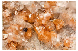
Hand Specimen Identification
Radioactivity, color, crystal habit, and associations help identify monazite. It may be confused with zircon, but is not as hard and has different forms. It can be distinguished from titanite by its crystal shape and high density.
Physical Properties
| hardness | 5 to 5.5 |
| specific gravity | 4.9 to 5.2 |
| cleavage/fracture | perfect (001), good (100)/subconchoidal |
| luster/transparency | variable, subresinous/translucent |
| color | red, brown, yellowish |
| streak | white |
Optical Properties
Monazite is colorless, gray, or yellow-brown in thin section. It has high positive relief and displays up to third or fourth order interference colors. Biaxial (+), α = 1.785 to 1.800 , β = 1.786 to 1.801, γ = 1.838 to 1.850, δ = 0.005, 2V = 10° to 20°.
Crystallography
Monazite is monoclinic, a = 6.79, b = 7.01, c = 6.46, β = 104.4°, Z = 4; space group \(P\dfrac{2_1}{n}\); point group \(\dfrac{2}{m}\).
Habit
Monazite crystals are usually small, tabular, or prismatic, often forming granular masses or individual grains in sand.
Structure and Composition
Monazite is isostructural with crocoite. In its structure, distorted (PO4)3- polyhedra are bonded to rare earth elements in 9-fold coordination. All the rare earths may be present, but Ce, La, and Th are usually the dominant large cations. Small amounts of Si may substitute for P in the tetrahedral sites.
Occurrence and Associations
Monazite is a rare secondary mineral is silicic igneous rocks. It is also found in unconsolidated beach or stream sediments, where it is associated with other heavy minerals such as magnetite and ilmenite.
Varieties
Rare earth content varies, so the names monazite-(Ce), monazite-(La), and so on are sometimes used to designate the dominant rare earth.
Related Minerals
Monazite is isostructural with crocoite, PbCrO4, and forms solid solutions with huttonite, ThSiO4. It is chemically related to xenotime, YPO4, with which it forms a minor solid solution.
Triphylite Li(Fe,Mn)PO4
Origin of Name
From the Greek words for “three” and “family,” in reference to its three cations.

Hand Specimen Identification
Brown-green to gray or bluish color, association with other pegmatite minerals, 90° cleavage angle, and resinous luster help identify triphylite.
Physical Properties
| hardness | 5 to 5.5 |
| specific gravity | 3.5 to 5.5 |
| cleavage/fracture | perfect (001), good (010)/subconchoidal |
| luster/transparency | vitreous, resinous/ transparent to translucent |
| color | variable brown-green to gray or bluish |
| streak | white, gray |
Optical Properties
Triphylite is biaxial (-), α = 1.68 , β = 1.68, γ = 1.69, δ = 0.01, 2V = 0° to 56°.
Crystallography
Triphylite is orthorhombic, a = 6.01, b = 4.68, c = 10.36, Z = 4; space group \(P\dfrac{2_1}{m}\dfrac{2_1}{c}\dfrac{2_1}{n}\); point group \(\dfrac{2}{m}\dfrac{2}{m}\dfrac{2}{m}\).
Habit
Euhedral crystals are rare; triphylite is typically fine grained and massive.
Structure and Composition
In triphylite, the cations occupy octahedra forming zigzag chains between (PO4)3- tetrahedra. A complete solid solution exists between the Fe and Mn end members. Compositions near the Mn end member are given the name lithiophilite.
Occurrence and Associations
Triphylite, typically a pegmatite mineral, is found with other phosphates, quartz, feldspar, spodumene, and beryl.
Apatite Ca5(PO4)3(OH,F,Cl)
Origin of Name
From the Greek word apate, meaning “deceit,” because it is often difficult to distinguish it from other minerals.
Hand Specimen Identification
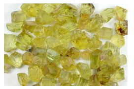
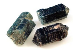
Apatite may be any of a number of different colors. The olive-green color seen in Figure 14.431 is most common. Other shades of green and blue are quite common, too, such as the blue color of the crystals in Figure 14.432. Other hues are relatively rare.
When euhedral, apatite crystals are easy to discern hexagonal prisms, often with terminating pyramidal faces. Cleavage traces run perpendicular to prism faces and long dimension. (Cleavage is quite obvious in Figure 14.432) Also aiding identification: apatite has moderate density, is softer than quartz and feldspar, but harder the calcite and fluorite.
Physical Properties
| hardness | 5 |
| specific gravity | 3.2 |
| cleavage/fracture | good (001), poor (100)/conchoidal |
| luster/transparency | subresinous/transparent to translucent |
| color | green, yellow, variable |
| streak | white |
Optical Properties
Apatite appears similar to quartz in thin section. It is colorless, does not develop good cleavage, and has very low birefringence. Quartz, however, has lower relief and is uniaxial (+). Apatite is uniaxial (-), ω = 1.633, ε = 1.630, δ = 0.003.
Crystallography
Apatite is hexagonal, a = 9.38, c = 6.86, Z = 2; space group \(P\dfrac{6_3}{m}\); point group \(\dfrac{6}{m}\).
Habit
Apatite typically forms prismatic crystals, but may be colloform, massive, or granular.
Structure and Composition
Complete solid solution exists between hydroxyapatite (OH end member), fluorapatite (F end member), and chlorapatite (Cl end member). In addition, transition metals or Sr2+ may replace Ca2+; and (CO3)2-, OH–, or (SO4)2- may replace some (PO4)3-. In the apatite structure, Ca-PO4 chains run parallel to the c-axis. Ca2+ is located around channels occupied by (F,Cl,OH).
Occurrence and Associations
Apatite is a common accessory mineral but only rarely a major rock former. It is common in all igneous rocks, including pegmatites and hydrothermal veins, in metamorphic rocks, and in marine sediments.
Varieties
Collophane is a massive cryptocrystalline form of apatite that comprises some phosphate rocks and bones.
Related Minerals
A large number of phosphates, sulfates, arsenates, vanadates, and silicates are isostructural with apatite, but none are common.
Pyromorphite Pb5(PO4)3Cl
Origin of Name
From the Greek words meaning “fire” and “form,” because it typically develops large faces when crystallizing from a magma.

Hand Specimen Identification
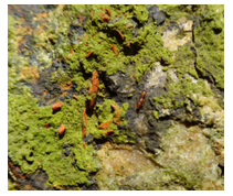
Occurrence with other Pb-minerals, bright green, green-yellow, or brown color, habit, high density, and sometimes resinous luster identify pyromorphite. It is sometimes confused with apatite, mostly because of its green color, but is softer than apatite.
Figure 14.433 shows green pyromorphite on gray galena (Pb-sulfide). Minor orange crocoite (Pb-chromate) can also be seen. The specimen in Figure 14.434 contains green pyromorphite and white/clear cerrusite (Pb-carbonate). The brown material around the edges may be crocoite.
Physical Properties
| hardness | 3.5 to 4 |
| specific gravity | 7.0 |
| cleavage/fracture | poor {100}, poor {101} |
| luster/transparency | resinous/transparent to translucent |
| color | bright green, yellow, variable |
| streak | white, yellow |
Optical Properties
Pyromorphite is uniaxial (-), ω = 2.058, ε = 2.048, δ = 0.010.
Crystallography
Pyromorphite is hexagonal, a = 9.97, c = 7.32, Z = 2; space group \(P\dfrac{6_3}{m}\); point group \(\dfrac{6}{m}\).
Habit
Pyromorphite is typically prismatic, having a barrel shape; it is less commonly granular, fibrous, cavernous (hollow prisms), globular, or reniform.
Structure and Composition
Pyromorphite is isostructural with apatite (see apatite structure). Some Ca may substitute for Pb. P may be replaced by As.
Occurrence and Associations
Pyromorphite is a secondary mineral, found with other oxidized Pb or Zn minerals, in oxidized zones associated with Pb deposits. It is isostructural with apatite and with mimetite, Pb(AsO4)3Cl, with which it forms a complete solid solution.
Amblygonite LiAl(PO4)F
Origin of Name
From the Greek word amblygonios, referring to its cleavage angle.

Hand Specimen Identification
Amblygonite is most often a nondescript white to creamy translucent mineral that is difficult to distinguish from other light-colored minerals with moderate density and hardness. Its single perfect cleavage and conchoidal fracture help identification and distinguish it from feldspars, feldspathoids, and zeolites. Its occurrence in pegmatites, especially if it contrasts with any feldspar present, also aids identification. Figure 14.435 shows typical nondescript amblygonite. Figure 10.64 (from an earlier chapter) shows an unusual yellow specimen.
Physical Properties
| hardness | 6 |
| specific gravity | 3.0 |
| cleavage/fracture | perfect (100), good (110), poor (011)/ subconchoidal |
| luster/transparency | vitreous, greasy/transparent to translucent |
| color | white, beige, green, sometimes other colors |
| streak | white |
Optical Properties
Amblygonite is biaxial (-), α = 1.59 , β = 1.60, γ = 1.62, δ = 0.03, 2V = 52° to 90°.
Crystallography
Amblygonite is triclinic, a = 6.644, b = 7.744, c = 6.91, α = 90.35°, β = 117.33°, γ = 91.01°, Z = 2; space group \(P\overline{1}\); point group \(\overline{1}\).
Habit
Rare crystals may be equant or columnar. Amblygonite is more commonly found as rough masses or in irregular aggregates.
Structure and Composition
Amblygonite is composed of alternating (PO4) tetrahedra and AlO5F octahedra linked by Li+ ions. F may be replaced by OH. Minor Na may be present.
Occurrence and Associations
Amblygonite is a rare mineral found in pegmatites rich in Li and P. Typical associated minerals include lepidolite, spodumene, apatite, and tourmaline.
Related Minerals
Several other phosphate minerals, some which can be gems, are similar to amblygonite and are found in pegmatites: herderite, CaBe(PO4)(F,OH); beryllonite, NaBePO4; and brazilianite, NaAl3(PO4)2(OH)4. Complete solid solution exists between amblygonite and a hydroxy end member, montebrasite, LiAl(PO4)(OH).
Lazulite (Mg,Fe)Al2(PO4)2(OH)2
Origin of Name
From an Arabic word meaning “heaven,” referring to its sky-blue color.

Hand Specimen Identification
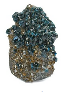
Lazulite’s azure-blue color is distinctive. Few minerals ever have a strong blue color like lazulite‘s. When crystals are visible, their pyramidal form distinguishes lazulite from the other blue minerals. When massive, identification is problematic.
Figure 14.436 shows typical lazulite crystals, up to several centimeters long, from Austria. The light blue color is typical. Some lazulite has a darker color; the photo of the specimen from the Yukon in Figure 14.437 shows an example.
Physical Properties
| hardness | 5 to 5.5 |
| specific gravity | 3.0 |
| cleavage/fracture | poor (011)/uneven |
| luster/transparency | vitreous/translucent |
| color | azure-blue |
| streak | white |
Optical Properties
Lazulite is biaxial (-), α = 1.612 , β = 1.634, γ = 1.643, δ = 0.031, 2V = 70°.
Crystallography
Lazulite is monoclinic, a = 7.16, b = 7.26, c = 7.24, β = 120.67°, Z = 2; space group \(P\dfrac{2_1}{c}\); point group \(\dfrac{2}{m}\).
Habit
Lazulite is typically massive, granular, or compact; rare crystals are prismatic or pyramidal with steep faces.
Structure and Composition
Octahedral Mg and Fe are linked to octahedral Al by sharing of O2- and OH–. The (Mg,Fe,Al)6 octahedra bond to (PO4)3- tetrahedra. Complete solid solution exists between the Mg and Fe end members. Scorzalite is the name of Fe-rich members of the series.
Occurrence and Associations
Lazulite and related minerals are rare, found only in some pegmatites and high-grade metamorphic rocks. They may be associated with rutile, kyanite, corundum, and sillimanite.
Wavellite Al3(PO4)2(OH)3•5H2O
Origin of Name
Named after W. Wavel (d. 1829), who discovered it.

Hand Specimen Identification
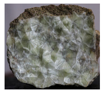
Wavellite is a secondary mineral that most commonly forms planar radiating clusters of crystals along fracture surfaces, creating “starburst” structures like those seen in Figure 14.438. Wavellite also forms globular radiating aggregates of acicular crystals that develop in voids. This produces spherical structures with a “cotton-ball” appearance, like the wavellite balls seen in Figure 14.439. The planar or cotton-ball radiating textures, and a light green to yellow color, are diagnostic for this mineral. Other occurrences are known, but not as easily identified as wavellite.
Physical Properties
| hardness | 3.5 to 4 |
| specific gravity | 2.36 |
| cleavage/fracture | perfect prismatic {101}, perfect (010)/subconchoidal |
| luster/transparency | vitreous/translucent |
| color | white, greenish yellow, gray, brown |
| streak | white |
Optical Properties
Wavellite is biaxial (+), α = 1.525 , β = 1.534, γ = 1.552, δ = 0.027, 2V = 72°.
Crystallography
Wavellite is orthorhombic, a = 9.62, b = 17.36, c = 6.99, Z = 4; space group \(P\dfrac{2_1}{c}\dfrac{2_1}{m}\dfrac{2_1}{n}\); point group \(\dfrac{2}{m}\dfrac{2}{m}\dfrac{2}{m}\).
Habit
2-dimensional or 3-dimensional radiating acicular crystal aggregates typify wavellite. Less commonly wavellite forms dense opal-like or stalactitic masses. Large visible individual crystals are very rare.
mo
Structure and Composition
Wavellite’s structure is incompletely known. It may contain small amounts of Fe and Mg.
Occurrence and Associations
Wavellite is a secondary mineral found in rock cavities or on joint surfaces in low-grade aluminous metamorphic rocks. It is also found in phosphorite deposits. Common associated minerals include other phosphate minerals and limonite.
Turquoise CuAl6(PO4)4(OH)8•4H2O
Origin of Name
From the French word turquoise, meaning “Turkish,” in reference to the source of the original stones imported into Europe.

Hand Specimen Identification
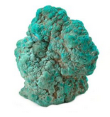
Turquoise forms in veins or as void fillings and is generally cryptocrystalline (exceptionally fine- grained), sometimes appearing amorphous. It typically has a distinctive turquoise-blue color seen in these two photos, but different shades of blue and green are well known. Its color and cryptocrystalline character generally serve for identification.
Turquoise is not often confused with other minerals. Chrysocolla, a blue-green hydrated Cu-silicate may have similar colors but is much softer. (Chrysocolla has hardness of 2-3 compared with turquoise‘s 6.) Malachite (green) and azurite (blue), both hydrated copper carbonates, have colors distinct from turquoise and hardnesses of 3.5 to 4.
Physical Properties
| hardness | 6 |
| specific gravity | 2.7 |
| cleavage/fracture | perfect but rarely seen (001), good (010)/subconchoidal, brittle |
| luster/transparency | resinous, waxy/translucent |
| color | turquoise and related blue-green shades, other hues are exceptionally rare |
| streak | white, green |
Optical Properties
Turquoise is biaxial (+), α = 1.61, β = 1.62, γ = 1.65, δ = 0.04, 2V = 40°.
Crystallography
Turquoise is triclinic, a = 7.48, b = 9.95, c = 7.69, α = 111.65°, β = 115.38°, γ = 69.43°, Z = 1; space group P1; space group P1; point group 1.
Habit
Rare small crystals of turquoise are known, but reniform, massive, or granular varieties are more typical.
Structure and Composition
The structure of turquoise consists of a framework of (PO4)3- tetrahedra and Al octahedra. Holes in the structure contain Cu, which bonds to the polyhedra, OH– and to H2O. Fe3+ may substitute for Al3+.
Occurrence and Associations
Turquoise occurs as a secondary mineral associated with Al-rich volcanic rocks. It forms in small seams, veins, stringers, and crusts. Associated minerals typically include kaolinite, Al2Si2O5(OH)4; limonite, Fe2O3•nH2O; or chalcedony, SiO2.
Varieties
Zn-rich blue-green varieties of turquoise are called faustite.
Related Minerals
Complete solid solution exists between turquoise and chalcosiderite, CuFe6(PO4)4(OH)8•4H2O.
Autunite Ca(UO2)2(PO4)2•10H2O
Origin of Name
Named for Autun, France, where it is found.


Hand Specimen Identification
A strong yellow or yellow-green color, radioactivity, fluorescence under ultraviolet light, and typically tetragonal platy crystals identify autunite.
Figure 14.442 shows autunite from France. Figure 14.443 show a reticulated cluster of autunite crystals from Portugal.
Physical Properties
| hardness | 2 to 2.5 |
| specific gravity | 3.15 |
| cleavage/fracture | perfect basal (001), good prismatic (100), (010) and {110 }/uneven |
| luster/transparency | pearly, adamantine/transparent to translucent |
| color | typically lemon yellow to greenish yellow to pale green |
| streak | yellow |
Optical Properties
Autunite is uniaxial (-), ω = 1.577, ε = 1.553, δ = 0.024.
Crystallography
Autunite is tetragonal, a = 7.00, c = 20.67, Z = 4; space group \(I\dfrac{4}{m}\dfrac{2}{m}\dfrac{2}{m}\); point group \(\dfrac{4}{m}\dfrac{2}{m}\dfrac{2}{m}\).
Habit
Thin tabular or flaky crystals, often square, are typical for autinite. It commonly forms reticulated masses of crusts.
Structure and Composition
In autunite, (PO4)3- tetrahedra link to U octahedra to form uneven layers. The layers are connected by weakly bonded H2O molecules. Other alkaline earths may substitute for Ca in small amounts; the amount of water in the structure is somewhat variable.
Occurrence and Associations
Autunite is a secondary uranium mineral, often forming after uraninite, UO2. Torbernite, Cu(UO2)2(PO4)2•nH2O, and uraninite are common associated minerals.
Related Minerals
Torbernite is isostructural with autunite and has similar properties.
Vanadinite Pb5(VO4)3Cl
Origin of Name
Named in reference to its vanadium content.


Hand Specimen Identification
Vanadinite generally has a distinctive bright red to orange-red color and forms hexagonal crystals. Color is the key to identification and distinguishes this mineral from apatite, pyromorphite, or mimetite. It may be transparent, translucent, or opaque and has an adamantine to resinous luster.
Figure 14.444 shows a typical occurrence of vanadinite with calcite from Mexico. Figure 14.445 is a photo of a spectacular museum specimen that originally came from Morocco. Enlarge this photo and the hexagonal nature of the crystals is easily seen.
Physical Properties
| hardness | 3 |
| specific gravity | 6.9 |
| cleavage/fracture | none/subconchoidal |
| luster/transparency | resinous/translucent |
| color | ruby red, orange-red, brown, yellow |
| streak | white, yellow |
Optical Properties
Vanadinite is uniaxial (-), ω = 2.416, ε = 2.350, δ = 0.066.
Crystallography
Vanadinite is hexagonal, a = 10.33, c = 7.35, Z = 2; space group \(P\dfrac{6_3}{m}\); point group \(\dfrac{6}{m}\).
Habit
Vanadinite often forms hexagonal prisms, sometimes hollow, with or without pyramidal faces. It may also form rounded or globular masses.
Structure and Composition
Vanadinite has the same structure as apatite (see apatite structure). P or As may substitute for V in small amounts. Minor amounts of Ca, Zn, and Cu may also be present.
Occurrence and Associations
Vanadinite is a rare mineral found in the oxidized portions of Pb deposits where it is often associated with galena, cerussite, or limonite.
Related Minerals
Vanadinite is isostructural with apatite, Ca5(PO4)3(OH,F,Cl), and with a number of other arsenates, vanadates, and phosphates. It forms solid solutions with mimetite, Pb5(AsO4)3Cl, and intermediate compositions are called endlichite.
Carnotite K2(UO2)2(VO4)2•3H2O
Origin of Name
Named after M. A. Carnot (1839–1920), a French mining engineer and Inspector General of Mines.


Hand Specimen Identification
Radioactivity and yellow color, often with a greenish stain, characterize carnotite. It is sometimes confused with other secondary uranium minerals.
Figure 14.446 shows typical fine-grained carnotite deposited in a sandstone. Individual crystals are exceptionally small. Figure 14.447 is a much magnified photo of carnotite crystals smaller than millimeter across.
Physical Properties
| hardness | 1 |
| specific gravity | 4.5 |
| cleavage/fracture | perfect but rarely seen (001)/uneven |
| luster/transparency | dull, earthy/translucent |
| color | yellow, yellow-green |
| streak | yellow |
Optical Properties
Carnotite is biaxial (-), α = 1.75 , β = 1.92, γ = 1.95, δ = 0.20, 2V = 38° to 44°.
Crystallography
Carnotite is monoclinic, a = 10.47, b = 8.41, c = 6.91, β = 103.67°, Z = 1; space group \(P\dfrac{2_1}{a}\); point group \(\dfrac{2}{m}\).
Habit
Fine powder or crumbly aggregates characterize carnotite. It may also be disseminated.
Structure and Composition
Carnotite‘s structure contains layers of edge-sharing uranium and vanadium polyhedra. The layers are joined by weak bonds to interlayer K and H2O.
Occurrence and Associations
Carnotite is a secondary uranium mineral typically found as crusts or flakes in sandstones or conglomerates that have been altered by circulation of meteoric waters.
Related Minerals
A number of other hydrated uranium oxides are known, including tyuyamunite, Ca(UO2)2(VO4)2•nH2O; torbernite, Cu(UO2)2(PO4)2•nH2O; and autunite, Ca(UO2)2(PO4)2 •10H2O.
Erythrite Co(AsO)2•8H2O
Origin of Name
From the Greek word erythros, meaning “red.”

Hand Specimen Identification
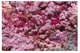
Erythrite is a secondary cobalt-arsenic mineral. It is one of only a few minerals that have its kind of red-pink-purple color; the two photos here show examples. Color, association with other cobalt minerals, and often crusty or drusy appearance generally identify this mineral.
Figure 14.448 is a photo of typical crusty erythrite. The specimen comes from Australia. Figure 14.449 shows a much enlarged view of a similar specimen from Morocco that contains both needles and cottony balls of erythrite. The largest needles and balls are several millimeters in longest dimension.
Physical Properties
| hardness | 1.5 to 2.5 |
| specific gravity | 3.06 |
| cleavage/fracture | perfect basal (010)/sectile |
| luster/transparency | adamantine/transparent to translucent |
| color | crimson, pink, purple-red |
| streak | pale purple |
Optical Properties
Erythrite is biaxial (-), α = 1.626, β = 1.661, γ = 1.699, δ = 0.073, 2V = 90°.
Crystallography
Erythrite is monoclinic, a = 10.26 , β = 13.37, c = 4.74, β = 105.1°, Z = 2; space group \(C\dfrac{2}{m}\); point group \(\dfrac{2}{m}\).
Habit
Large, easily seen crystals are rare. Erythrite is typically in prismatic, acicular, reniform, or globular groups/clusters. Erythrite typically forms as drusy coatings and crusts and may be earthy or powdery.
Structure and Composition
The atomic structure in erythrite is layered, with vertex sharing by As tetrahedra and Co octahedra. Ni may substitute for Co.
Occurrence and Associations
Erythrite may form as a pink powdery coating, called cobalt bloom, on other cobalt minerals such as cobaltite, (Co,Fe)AsS, or skutterudite, (Co,Ni)As3.
Related Minerals
Annabergite, Ni3(AsO4)2•8H2O, also called nickel bloom, is isostructural with erythrite, but has an apple-green color. A complete solid solution exists between erythrite and annabergite.


