8.2: Diagenetic Structures
- Page ID
- 20962
\( \newcommand{\vecs}[1]{\overset { \scriptstyle \rightharpoonup} {\mathbf{#1}} } \)
\( \newcommand{\vecd}[1]{\overset{-\!-\!\rightharpoonup}{\vphantom{a}\smash {#1}}} \)
\( \newcommand{\dsum}{\displaystyle\sum\limits} \)
\( \newcommand{\dint}{\displaystyle\int\limits} \)
\( \newcommand{\dlim}{\displaystyle\lim\limits} \)
\( \newcommand{\id}{\mathrm{id}}\) \( \newcommand{\Span}{\mathrm{span}}\)
( \newcommand{\kernel}{\mathrm{null}\,}\) \( \newcommand{\range}{\mathrm{range}\,}\)
\( \newcommand{\RealPart}{\mathrm{Re}}\) \( \newcommand{\ImaginaryPart}{\mathrm{Im}}\)
\( \newcommand{\Argument}{\mathrm{Arg}}\) \( \newcommand{\norm}[1]{\| #1 \|}\)
\( \newcommand{\inner}[2]{\langle #1, #2 \rangle}\)
\( \newcommand{\Span}{\mathrm{span}}\)
\( \newcommand{\id}{\mathrm{id}}\)
\( \newcommand{\Span}{\mathrm{span}}\)
\( \newcommand{\kernel}{\mathrm{null}\,}\)
\( \newcommand{\range}{\mathrm{range}\,}\)
\( \newcommand{\RealPart}{\mathrm{Re}}\)
\( \newcommand{\ImaginaryPart}{\mathrm{Im}}\)
\( \newcommand{\Argument}{\mathrm{Arg}}\)
\( \newcommand{\norm}[1]{\| #1 \|}\)
\( \newcommand{\inner}[2]{\langle #1, #2 \rangle}\)
\( \newcommand{\Span}{\mathrm{span}}\) \( \newcommand{\AA}{\unicode[.8,0]{x212B}}\)
\( \newcommand{\vectorA}[1]{\vec{#1}} % arrow\)
\( \newcommand{\vectorAt}[1]{\vec{\text{#1}}} % arrow\)
\( \newcommand{\vectorB}[1]{\overset { \scriptstyle \rightharpoonup} {\mathbf{#1}} } \)
\( \newcommand{\vectorC}[1]{\textbf{#1}} \)
\( \newcommand{\vectorD}[1]{\overrightarrow{#1}} \)
\( \newcommand{\vectorDt}[1]{\overrightarrow{\text{#1}}} \)
\( \newcommand{\vectE}[1]{\overset{-\!-\!\rightharpoonup}{\vphantom{a}\smash{\mathbf {#1}}}} \)
\( \newcommand{\vecs}[1]{\overset { \scriptstyle \rightharpoonup} {\mathbf{#1}} } \)
\(\newcommand{\longvect}{\overrightarrow}\)
\( \newcommand{\vecd}[1]{\overset{-\!-\!\rightharpoonup}{\vphantom{a}\smash {#1}}} \)
\(\newcommand{\avec}{\mathbf a}\) \(\newcommand{\bvec}{\mathbf b}\) \(\newcommand{\cvec}{\mathbf c}\) \(\newcommand{\dvec}{\mathbf d}\) \(\newcommand{\dtil}{\widetilde{\mathbf d}}\) \(\newcommand{\evec}{\mathbf e}\) \(\newcommand{\fvec}{\mathbf f}\) \(\newcommand{\nvec}{\mathbf n}\) \(\newcommand{\pvec}{\mathbf p}\) \(\newcommand{\qvec}{\mathbf q}\) \(\newcommand{\svec}{\mathbf s}\) \(\newcommand{\tvec}{\mathbf t}\) \(\newcommand{\uvec}{\mathbf u}\) \(\newcommand{\vvec}{\mathbf v}\) \(\newcommand{\wvec}{\mathbf w}\) \(\newcommand{\xvec}{\mathbf x}\) \(\newcommand{\yvec}{\mathbf y}\) \(\newcommand{\zvec}{\mathbf z}\) \(\newcommand{\rvec}{\mathbf r}\) \(\newcommand{\mvec}{\mathbf m}\) \(\newcommand{\zerovec}{\mathbf 0}\) \(\newcommand{\onevec}{\mathbf 1}\) \(\newcommand{\real}{\mathbb R}\) \(\newcommand{\twovec}[2]{\left[\begin{array}{r}#1 \\ #2 \end{array}\right]}\) \(\newcommand{\ctwovec}[2]{\left[\begin{array}{c}#1 \\ #2 \end{array}\right]}\) \(\newcommand{\threevec}[3]{\left[\begin{array}{r}#1 \\ #2 \\ #3 \end{array}\right]}\) \(\newcommand{\cthreevec}[3]{\left[\begin{array}{c}#1 \\ #2 \\ #3 \end{array}\right]}\) \(\newcommand{\fourvec}[4]{\left[\begin{array}{r}#1 \\ #2 \\ #3 \\ #4 \end{array}\right]}\) \(\newcommand{\cfourvec}[4]{\left[\begin{array}{c}#1 \\ #2 \\ #3 \\ #4 \end{array}\right]}\) \(\newcommand{\fivevec}[5]{\left[\begin{array}{r}#1 \\ #2 \\ #3 \\ #4 \\ #5 \\ \end{array}\right]}\) \(\newcommand{\cfivevec}[5]{\left[\begin{array}{c}#1 \\ #2 \\ #3 \\ #4 \\ #5 \\ \end{array}\right]}\) \(\newcommand{\mattwo}[4]{\left[\begin{array}{rr}#1 \amp #2 \\ #3 \amp #4 \\ \end{array}\right]}\) \(\newcommand{\laspan}[1]{\text{Span}\{#1\}}\) \(\newcommand{\bcal}{\cal B}\) \(\newcommand{\ccal}{\cal C}\) \(\newcommand{\scal}{\cal S}\) \(\newcommand{\wcal}{\cal W}\) \(\newcommand{\ecal}{\cal E}\) \(\newcommand{\coords}[2]{\left\{#1\right\}_{#2}}\) \(\newcommand{\gray}[1]{\color{gray}{#1}}\) \(\newcommand{\lgray}[1]{\color{lightgray}{#1}}\) \(\newcommand{\rank}{\operatorname{rank}}\) \(\newcommand{\row}{\text{Row}}\) \(\newcommand{\col}{\text{Col}}\) \(\renewcommand{\row}{\text{Row}}\) \(\newcommand{\nul}{\text{Nul}}\) \(\newcommand{\var}{\text{Var}}\) \(\newcommand{\corr}{\text{corr}}\) \(\newcommand{\len}[1]{\left|#1\right|}\) \(\newcommand{\bbar}{\overline{\bvec}}\) \(\newcommand{\bhat}{\widehat{\bvec}}\) \(\newcommand{\bperp}{\bvec^\perp}\) \(\newcommand{\xhat}{\widehat{\xvec}}\) \(\newcommand{\vhat}{\widehat{\vvec}}\) \(\newcommand{\uhat}{\widehat{\uvec}}\) \(\newcommand{\what}{\widehat{\wvec}}\) \(\newcommand{\Sighat}{\widehat{\Sigma}}\) \(\newcommand{\lt}{<}\) \(\newcommand{\gt}{>}\) \(\newcommand{\amp}{&}\) \(\definecolor{fillinmathshade}{gray}{0.9}\)Admittedly there is no clear temporal cut-off between the secondary structures described in Chapter 4 (soft-sediment deformation structures, mudcracks, etc.) and the diagenetic structures list below - except to say that the diagenetitc structures generally form somewhat later and might be more disconnected from the processes that were active when the sediment was being deposited.
Diagenetic features formed by the growth of minerals/cement
Concretions are concentric accumulations of mineral cement. They come in a wide variety of shapes and sizes. Rhizoconcretions (aka rhizoliths) are a specific type of concretion developed around roots and commonly made of calcite or siderite.
Nodules are similar to concretions, except that they are internally massive. Distinctive types include:
- Caliche nodules – composed of calcite formed in association with soil forming processes
- Bauxites and laterites – nodular soils composed of iron- and alumina-rich minerals that form in response to intense tropical weathering.
- Septarian nodules – nodules with radial or concentric cracks, commonly filled with calcite or siderite crystals
- Desert rose – although it does not exactly fit the definition of a nodule, it is a rose- or flower shaped accumulation of gypsum
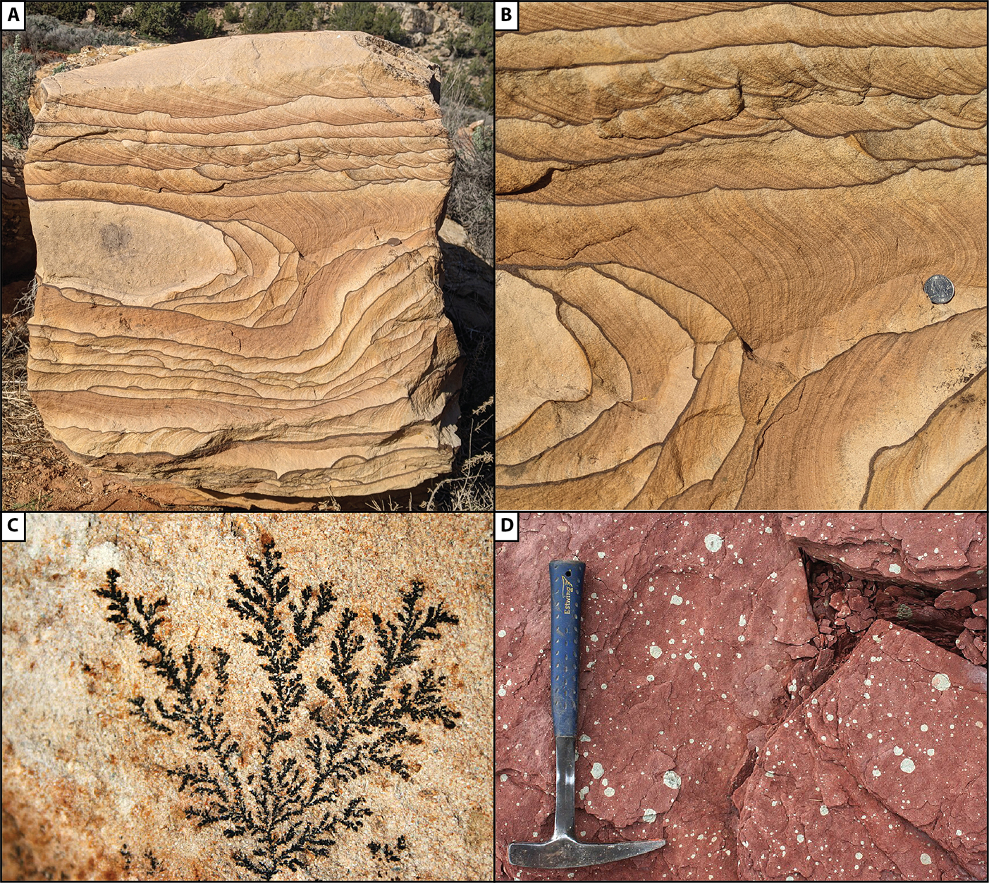
Liesegang rings – colored bands that formed as minerals precipitated from groundwater; commonly cut across bedding and may form elaborate curved patterns.
Manganese dendrites – chemical precipitate of MnO2 that forms on rock faces or bedding planes as a result of subaerial weathering. Commonly takes on a fern- or plant-like appearance.
Mottling – zones of distinctive color within rocks formed as a result of geochemical variations within the rock. May develop around organic-rich zones or areas with differences in porosity/permeability. Some may form circular or spherical reduction halos.
Features associated with dissolution
Geodes – layered accumulations of minerals that infill voids. They can form in voids of any origin and commonly form in cavities formed by dissolution of carbonates
Geopetal structures - small cavities formed beneath shells, etc. that contain crystals that are in the formerly open spaces. Useful for determining way up.
Secondary porosity - pores develop from the removal of material; commonly by selectively targeting certain grains or fossils.
Stylolites – irregular, jagged lines caused by pressure solution and the accumulation of insoluble residues.
Structures formed at the surface
Pedogenic slickensides – polished surfaces in mudrock formed in response shrinking and swelling associated with soil-forming processes. A hallmark of Vertisols.
Bioturbation – tracks, trails, burrows, or any feature formed from the activity of organisms.
Recrystallization structures
Cone-in-cone structures - zones within limestones that have fibrous calcite crystals that form a zig-zag pattern. They form in response to compaction and recrystallization.
Pseudospar – Coarsely crystalline calcite formed from the recrystallization of fine-grained lime mud (micrite).
Replacement features
Dolomitization – calcite/aragonite is replaced by dolomite. Dolomite shows up as rhombohedral crystals in thin section.
Replaced fossils (or other material) - forms when silica, pyrite, or some other mineral replacement occurs. Carbonate skeletons are prone to replacement.
Chickenwire structures – distinctive nodular zones in evaporates, commonly formed in response to the transition from gypsum to anhydrite and the incorporation of clastic sediment.
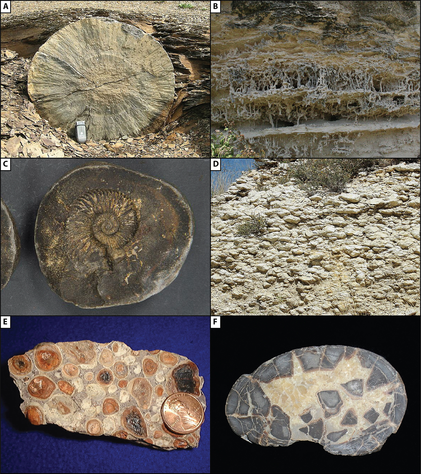
Figure \(\PageIndex{1}\): Diagnetic features formed in association with the growth of minerals or cement, part 1. A) Cross-section through a concretion. Although this example has a striking radial fabric, you can still see faint concentric layering (Mark Buchanan via Wikimedia Commons; CC BY-SA 3.0). B) Vertical rhizoconcretions developed around roots (Jfoote via Wikimedia Commons; CC BY-SA 3.0). C) Internally massive nodule developed around a coiled cephalopod (Hannes Grobe - AWI via Wikimedia Commons; CC BY-SA 4.0). D) Dense accumulation of modern caliche nodules in Texas (David R. Tribble via Wikimedia Commons; CC BY-SA 3.0). E) Nodules in a sample of bauxite (USGS via Wikimedia Commons; public domain). F) Septarian nodule with distinctive cracked texture (Neptunerover via Wikimedia Commons; public domain).

« Previous page
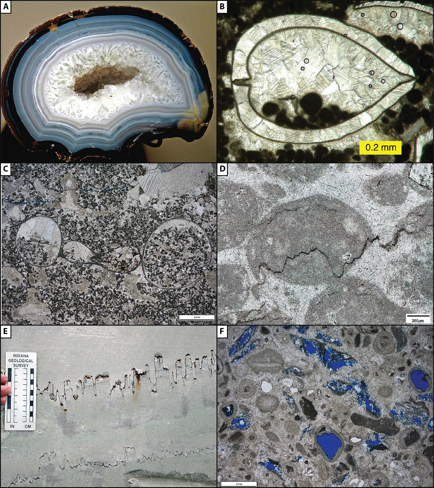
Figure \(\PageIndex{4}\): Diagenetic features formed in association with dissolution. A) Geode infilled with quartz crystals (James St. John via Wikimedia Commons; CC BY 2.0). B) Thin-section image of peloids and calcite cement inside of a recrystallized bivalve shell. The peloids settled to the bottom of the shell before the calcite was precipitated and thus are geopetal structures showing that up was toward the top of the page (Mark Wilson via Wikimedia Commons; public domain). C) Similar geopetal structure formed by sediment inside of a coiled gastropod or cephalopod shell (Michael C. Rygel via Wikimedia Commons; CC BY -SA 4.0). D & E show stylolites in thin section and in outcrop, respectively. F) Moldic porosity in a limestone formed by the selective dissolution of ooids. D-F from Michael C. Rygel via Wikimedia Commons; CC BY -SA 4.0.
Next page »
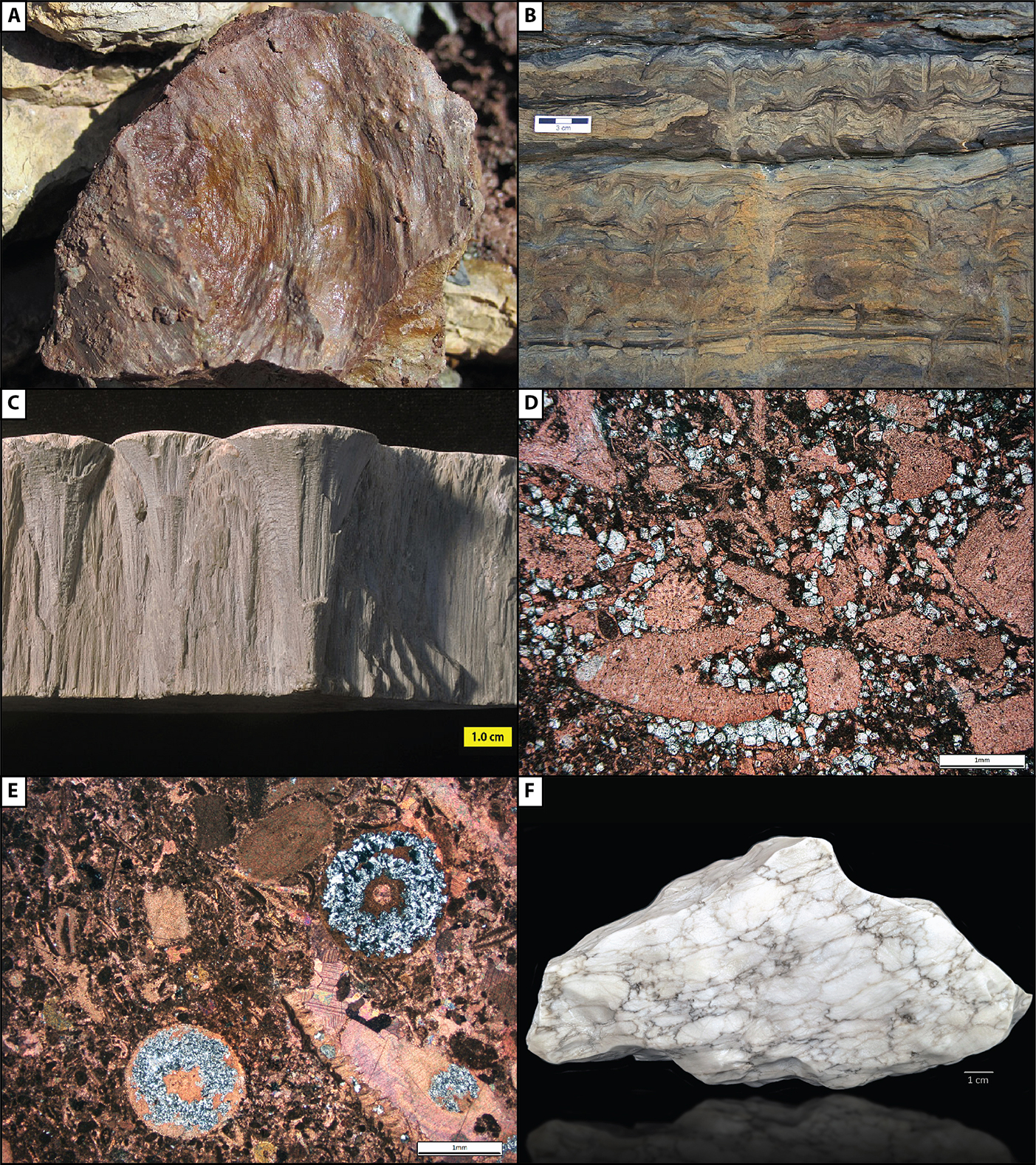
Figure \(\PageIndex{5}\): Diagenetic features formed at the surface, in association with recrystallization (A-B) or replacement (C-F). A) Pedogenic slickensides in a Mississippian paleosol (James St. John via Wikimedia Commons; CC BY 2.0). B) Trace fossils in Permian-aged shallow marine/deltaic sediments. C) Cone-in-cone limestone (Mark Wilson via Wikimedia Commons; public domain). D) Partially dolomitized fossiliferous limestone (PPL). Slide was stained for calcite which makes dolomite crystals (white) stand out from the fossils (light red) and micrite (dark reddish brown). E) Photomicrograph (XPL) of partially silicified fossils in limestone; slide was stained for calcite. F) Gypsum and anhydrite showing "chicken wire" texture. B, D, E, and F from Michael C. Rygel
