
5.4.1: The Components of a Microscope
- Page ID
- 19126

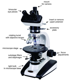
Polarizing microscopes, like the one seen in Figure 5.22, are in many respects the same as other microscopes. They magnify small features in a thin section so we can see fine details. These microscopes include many components. We view thin sections in two modes, depicted in Figure 5.23.
Orthoscopic illumination is standard and by far the most commonly used method. It involves an unfocused light beam that travels from the substage, through the thin section, and straight up the microscope tube to the ocular lens and our eyes. The light rays travel perpendicular to the stage and perpendicular to a thin section on the stage.
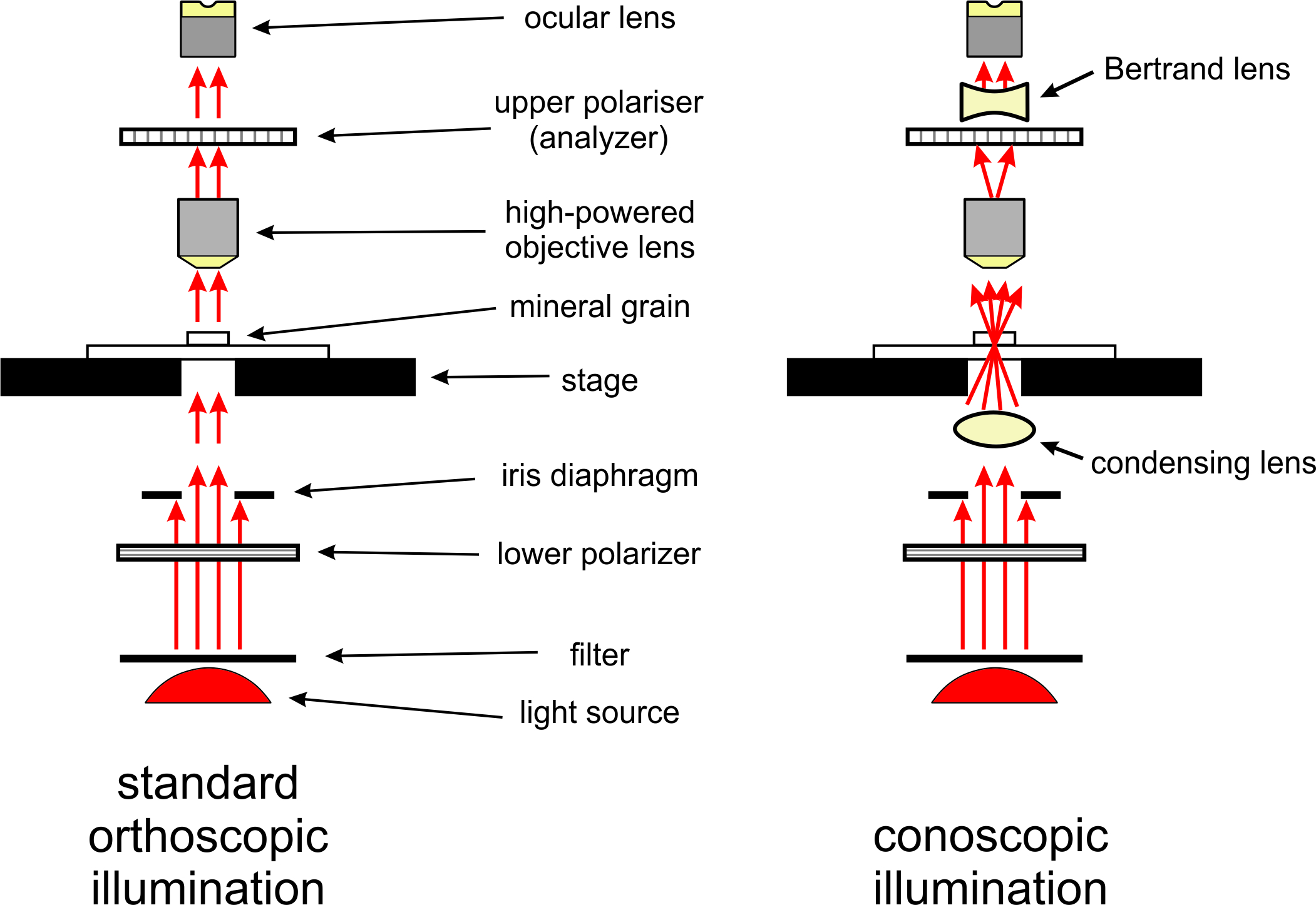
For some purposes, we insert a special lens called, a conoscopic lens, between the lower polarizer and stage to produce conoscopic illumination (also shown in Figure 5.23). The conoscopic lens, also called a condenser lens, causes the light beam to converge (focus) on a small spot on the sample. So, light illuminates the sample with a cone of nonparallel rays. The light then travels up the microscope tube in many directions instead of only vertically. Above the upper polarizer, most microscopes have a Bertrand lens that causes light to again travel vertically before it reaches the ocular.
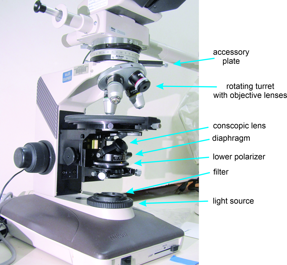
Figures 5.22 and 5.24 show the most important microscope components. A bulb beneath the microscope stage provides a white light source. The light passes through several filters, the lower polarizer, and a diaphragm that can limit the size of the light beam. When polarized white light reaches the stage, it interacts with the material being observed. Ultimately, the light reaches our eye(s) and we see the sample.
The most important filter below the stage is the lower (substage) polarizer, which ensures that all light striking the sample is plane polarized (vibrating, or having wave motion, in only one plane). The presence of a substage polarizer sets petrographic/polarizing microscopes apart from other microscopes. In most modern polarizing microscopes, the lower polarizer only allows light vibrating in an east-west direction to reach the stage. Older microscopes, however, may have the lower polarizer oriented in a north-south direction. Above the polarizing filter, a diaphragm helps concentrate light on the sample.
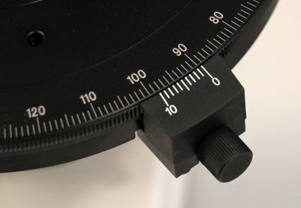
Because most minerals are anisotropic, the interaction of the light with a mineral varies with stage rotation. We can rotate the microscope stage to change the orientation of the sample relative to the polarized light. A calibrated angular scale around the outside of the stage allows us to make precise measurements of crystal orientation (Figure 5.25). The scale is also useful for measuring angles between cleavages, crystal faces, and twin orientations, and for measuring other optical properties.
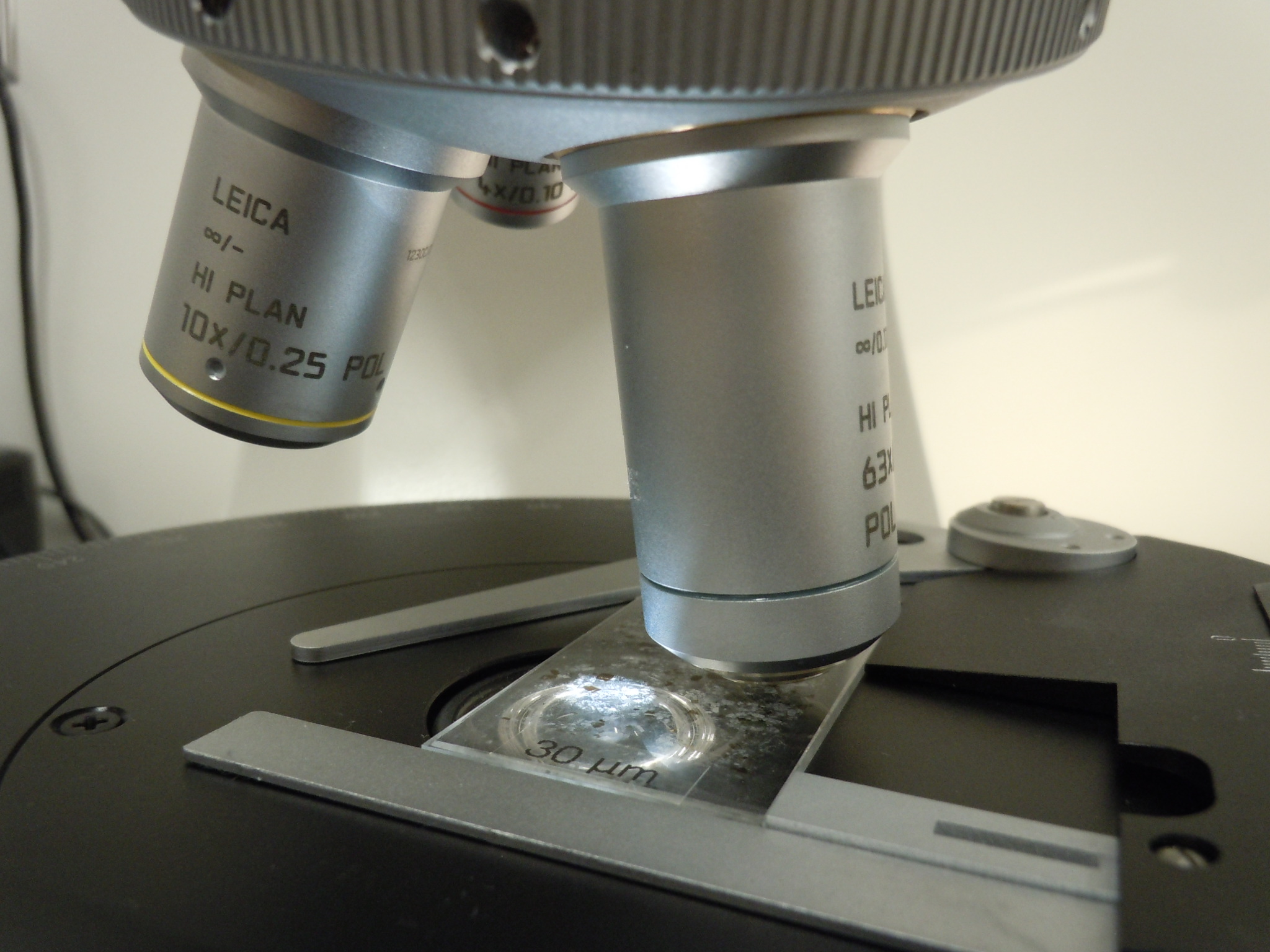
A rotating turret above the microscope stage holds several objective lenses (Figure 5.26). Typically, they range in magnification from 4x to 64x. Different objective lenses can have different numerical apertures (NA), a value that describes the angles at which light can enter a lens, which is an important consideration when making some kinds of measurements. In the discussion of interference figures later in this chapter, we have assumed that the objective lens being used has an NA of 0.85, since this is by far the most common today. (For lenses with a different NA, some angular values in the discussion of interference figures later in this chapter will be in error.)
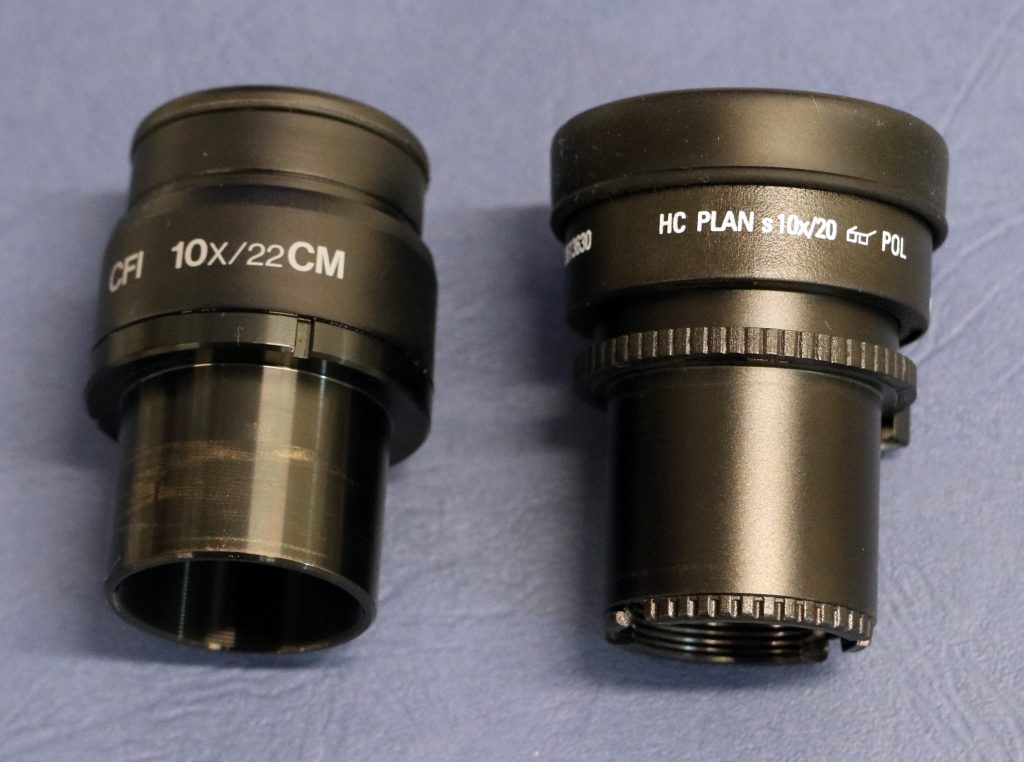
Microscope eyepieces contain additional lenses, called oculars, usually providing 8x or 10x magnification. (The eyepieces shown in Figure 5.27 have 10x magnification written on them.) Older microscopes only had one eyepiece but today most are binocular microscopes with two eyepieces and two oculars. Oculars have crosshairs that aid in making measurements when we rotate the stage. The total magnification, which is the product of the objective lens magnification and the ocular magnification, varies from about 16x to 500x, depending on the lenses used.
Petrographic microscopes have other filters and lenses between the objective lens and the ocular (shown and labeled in Figures 5.22, 5.23, and 5.24). The upper polarizer, sometimes called the analyzer, is a polarizing filter oriented at 90° to the lower polarizer. We can insert or remove it from the path of the light beam. If no sample is on the stage, light that passes through the lower polarizer cannot pass through the upper polarizer. If a sample is on the stage, it usually changes the polarization of the light so that some can pass through the upper polarizer.
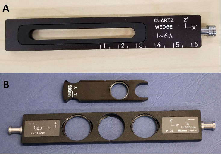
We can also insert a quartz wedge or an accessory plate below the upper polarizer. (A quartz wedge is exactly what it sounds like – a sliver of quartz that is thicker at one end than at the other.) The most common kind of accessory plate used today is a full wave plate. In the past, all full-wave plates were made of gypsum but today they are made of quartz. A wedge or an accessory plate modify light properties for making some specific kinds of observations.